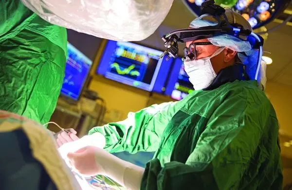
A 10-second intraoperative question
With an international team of collaborators, Hollon developed FastGlioma, an AI-powered system designed to answer a critical question in about 10 seconds:
Is tumor still present in tissue that could be safely removed?
In a study published in Nature, FastGlioma was found to detect and calculate how much tumor remained with about 92% average accuracy using full-resolution imaging. A rapid “fast mode” delivered results in roughly 10 seconds at about 90% accuracy.
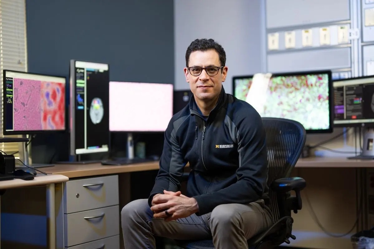
“FastGlioma is an artificial intelligence-based diagnostic system that has the potential to change the field of neurosurgery by immediately improving comprehensive management of patients with diffuse gliomas,” Hollon explained.
Why tumors can still be missed
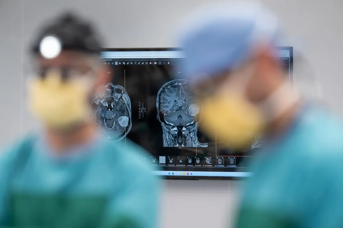
Even with modern tools, residual tumor can be hard to spot. Tumor infiltration and normal brain tissue can look nearly identical at the resection margin.
Some centers use intraoperative MRI, but it requires specialized equipment that isn’t available everywhere. Fluorescent imaging agents can help highlight certain tumors, but they can’t be applied to all tumor types.
The result is a persistent gap between what surgeons can see and what they need to know. Over the last 20 years, rates of residual tumor after neurosurgery have not meaningfully improved, and leaving tumor behind can contribute to worse outcomes.
FastGlioma is designed to close that gap at the exact moment decisions are made.
How FastGlioma performed
In comparison with conventional image- and fluorescent-guided approaches, FastGlioma missed high-risk residual tumor only 3.8% of the time, compared with nearly 25% for conventional methods.
“This means that we can detect tumor infiltration in seconds with extremely high accuracy, which could inform surgeons if more removal is needed during an operation,” Hollon said.
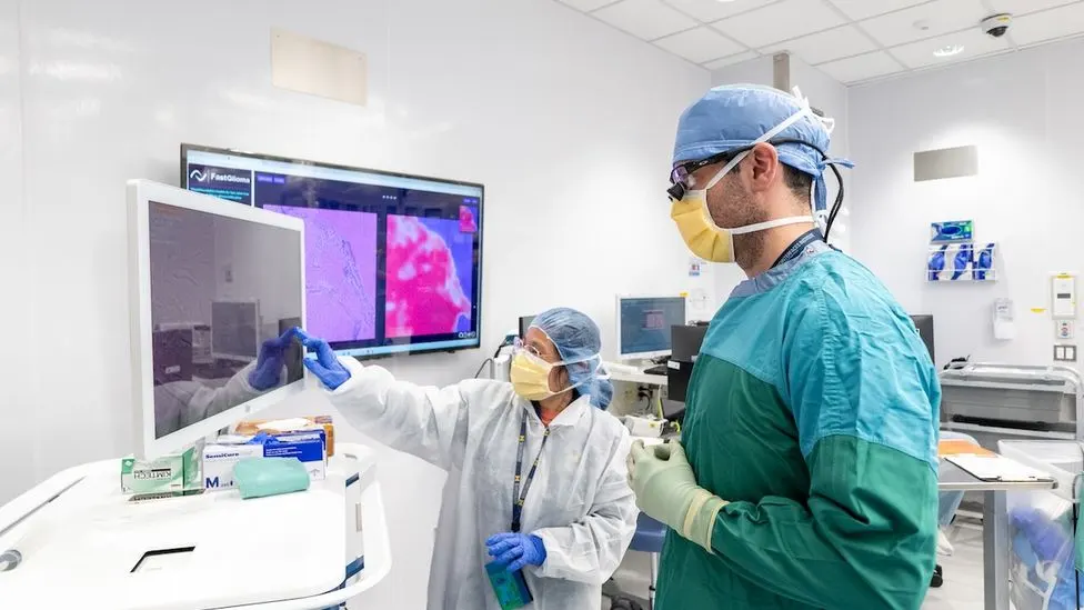
How it works: imaging + foundation models
FastGlioma combines rapid microscopic imaging with an AI approach known as foundation models—systems trained on large, diverse datasets that can be adapted across tasks.
The imaging foundation is based on a U‑M-developed technique called stimulated Raman histology.
“It’s a fancy name, but really all it means is that you can take a piece of tissue and you can put it in a microscope and you’re going to get very good images out… so good that you can see individual cells,” Hollon explained.
Historically, images of this quality were “way out of our reach” to obtain within seconds in an operating room.
Once those images could be captured during surgery, Hollon says, “we could use those images to train AI models… to make decisions about diagnosis of the tumor, whether this was tumor-infiltrated tissue”—in service of better brain tumor surgeries.
Built for real-time decisions
Full-resolution stimulated Raman histology images take around 100 seconds to acquire.
For real-time surgical guidance, the team also developed a lower-resolution “fast mode” that takes about 10 seconds, enabling near-immediate feedback in the OR.
To build FastGlioma, investigators pre-trained the model using more than 11,000 surgical specimens and 4 million unique microscopic fields of view, helping the system learn subtle patterns of tumor infiltration at scale.
Why seconds matter in glioma surgery
Gliomas were a central focus because outcomes are closely tied to how much tumor can be safely removed.
“Survival is really a function of how much tumor we remove and how well we do as brain tumor surgeons,” Hollon said.
“The reason it’s so important to know during surgery, within seconds, whether we have removed as much of the tumor as we safely can,” Hollon explained, “is because we want to make sure that all of the tumor that we can safely remove is in fact removed… [and] we want to make sure that if we’re getting close to an eloquent area in the brain—an area responsible for language function or motor function—we certainly don’t want to be removing any healthy portions of the brain.”
Tested beyond Michigan
The team didn’t just want a tool that worked at Michigan.
The study included a multicenter collaboration with partners at New York University, the University of California, San Francisco, and the Medical University of Vienna, helping to demonstrate that the approach can generalize across institutions and patient populations.
“We were able to show that not only can FastGlioma help patients here at the University of Michigan, but it works across the globe,” Hollon said.
Part of a broader vision for clinician-centered AI
FastGlioma is one piece of a larger effort to build AI tools across the care journey—before, during, and after surgery.
Hollon’s team at the Machine Learning in Neurosurgery Lab at U‑M is also developing diagnostic tools for neurological imaging to streamline triage and treatment for head trauma and other neurological conditions.
DeepGlioma: molecular diagnosis in under 90 seconds
One related tool, DeepGlioma, accelerates molecular diagnosis by analyzing tumor genetics in under 90 seconds during surgery.
For patients with diffuse glioma, among the deadliest brain tumors, speed can enable faster, more accurate classification and prognostic information to help guide surgical decisions and treatment plans. DeepGlioma could also support clinical trial enrollment, making targeted therapies more accessible.
Prima: MRI reads and triage in seconds
The team’s latest model, Prima, reads and diagnoses brain MRIs in seconds.
Tested on more than 30,000 MRI studies, Prima achieved up to 97.5% accuracy across more than 50 neurological disorders, outperforming other state-of-the-art models.
It can also automatically prioritize urgent cases, alerting specialists—such as stroke neurologists or neurosurgeons—for rapid intervention. Prima’s innovation lies in integrating patient medical history, imaging, and physician notes, mimicking a radiologist’s comprehensive approach and promising to streamline workflows throughout healthcare—even in resource-limited settings.
Together, FastGlioma, DeepGlioma, and Prima point toward a future where AI can strengthen clinical judgment with faster, more consistent information—supporting doctors and care teams in providing the best patient care possible.
Going beyond diffuse gliomas
FastGlioma can also be used to detect residual tumor for several non-glioma diagnoses, including pediatric brain tumors (such as medulloblastoma and ependymoma) and meningiomas.
Future studies will explore applying the workflow to other cancers, including lung, prostate, breast, and head and neck.
For Hollon, the long-term goal is global: “One day I do hope to see FastGlioma being used for every tumor surgery around the world.”